Services: Imaging
Testing and diagnostic procedures are integral in the identification and treatment of orthopedic and sports medicine conditions. The outstanding staff at the Orthopedic Center of Florida (OCF) uses advanced technology to ensure the highest quality of your diagnostic images. Our mission is to provide our patients with a compassionate treatment plan and offer comprehensive services needed for state of the art treatment with an emphasis on early activity and minimum down time.

Digital X-Ray
A proper X-Ray is probably the first and foremost piece of diagnostic information that aides in your diagnosis and treatment. OCF’s X-Ray department is completely digital, offering the following:
Immediate Availability – The procedure is usually completed within ten minutes. All images are immediately stored in PACS (Picture Archiving Communication System) and sent to your doctor’s computer.
High Quality & Resolution with Low Radiation – The level of radiation exposure from X-Rays is not harmful, but we will take special precautions if a patient is pregnant.
Unique Protocols – Limb length discrepancy evaluation is offered to create an exact template of lower limb lengths, which assist in treating malalignment and is used for surgical planning in the lower extremity
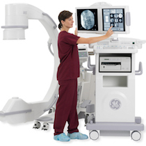
Fluoroscopy
Fluoroscopy is an imaging technique that uses low dose focused x-rays for guidance during injections and procedures. This imaging modality allows for enhanced visualization and more accurate placement of injections with less trauma and the ability to use smaller instruments.
Fluoroscopy can be used in conjunction with certain injectable dyes that enhance visualization and improve diagnosis. It can also be used for real time fracture reduction when manipulation and correction is needed in the office.
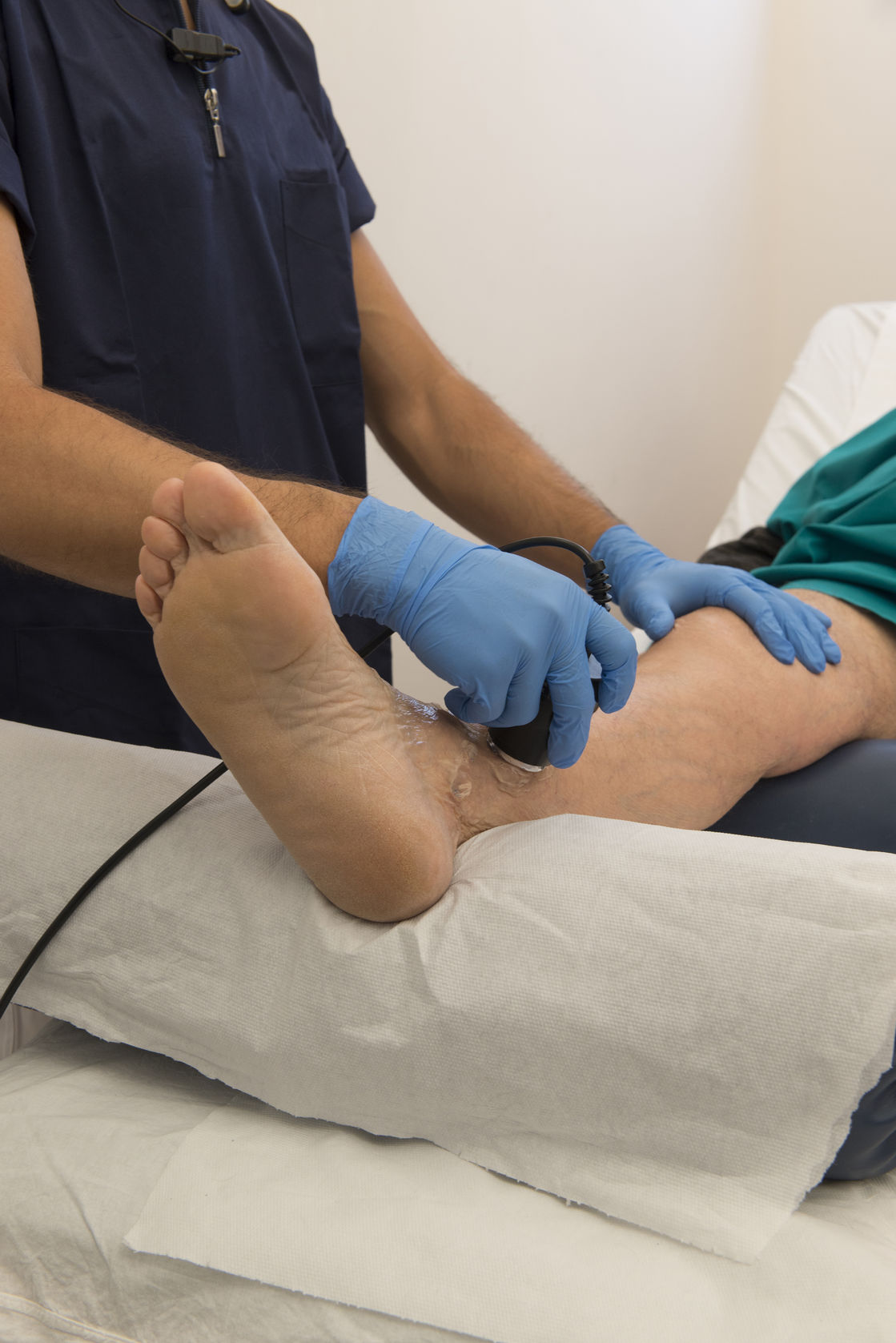
Musculoskeletal Ultrasound
High resolution ultrasound can be used as a first line imaging modality to assess and diagnose soft tissue pathology of tendons, muscles, ligaments, and peripheral nerves of the musculoskeletal system.
Ultrasound benefits include:
- Real time evaluation of soft tissue thickness, viscosity of fluid, and point of maximal intensity pain.
- Ability to evaluate structures through range of motion.
- Evaluation of stress fractures, joint inflammation, strains/sprains, tendonitis, hematoma, muscle tears, and disruptions.
- Provides PRECISE guidance for injections, needle placement for biopsy, aspiration, and visualize of needle placement.
- Non-invasive, cost effective modality that frequently eliminated the necessity for an MRI.
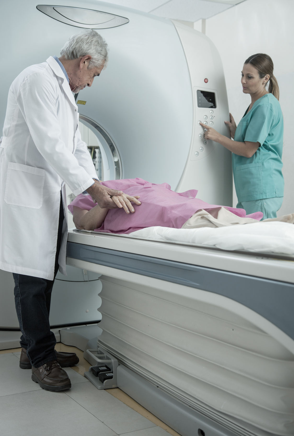
Open Extremity MRI
Many times a physician needs more information than a regular x-ray can demonstrate; that’s when an MRI is indicated. An MRI (Magnetic Resonance Imaging) is unlike an X-Ray as it contains no radiation, it uses magnets and radio waves to produce detailed images of bones, ligaments, tendons, cartilage and all surrounding soft tissue. The procedure at OCF includes:
An Appointment: Each MRI requires an appointment and takes approximately 30 to 90 minutes. Because of the strong magnetic field, we cannot perform MRI’s on patients with pacemakers or implanted aneurysm clips in the brain, or if you are pregnant.
Imaging: The patient is positioned on the open MRI table and should remain as motionless as possible. The MRI creates a magnetic field, and then pulses radio waves to the area of your body to be pictured. The radio waves cause your tissues to resonate. A computer records the rate at which your body’s various parts (tendons, ligaments, nerves, etc.) give off these vibrations, and translates the data into a detailed, two-dimensional picture. You will not feel any pain while undergoing an MRI, but the machine may be noisy.
Safety: You will be asked to fill out a questionnaire prior to your test and will also be asked to remove anything and everything that may be affected by the magnet such as cell phones, car keys, credit cards, jewelry, belts, watches, hair clips, money clips, glasses, hearing aids, etc. Once the scan is finished, beautiful, diagnostic images are sent and stored on our PACS (Picture Archiving Communication System) for the physician to review and discuss with you at your next clinic appointment.